


- Characteristics of Piezoelectric Polymeric PVDF Sensor by Impact Testing
Sojeong Kim, Ji-Yeon Shin, Yonghyeon Yun, Deuk Yong Lee†
 , Dong-Ho Yang*, Bae-Yeon Kim**, and Yo-Seung Song***
Department of Biomedical Engineering, Daelim University, Anyang 13916, Korea
*Control Factory Co., Ansan 15431, Korea
**Department of Materials Science and Engineering, Incheon National University, Incheon 22012, Korea
***Department of Materials Engineering, Korea Aerospace University, Goyang 10540, Korea- 충격시험을 이용한 압전고분자 PVDF 센서의 특성
김소정 · 신지연 · 윤용현 · 이득용†
· 양동호* · 김배연** · 송요승***대림대학교 의공융합과, *㈜Control Factory, **인천대학교 신소재공학과, ***한국항공대학교 재료공학과
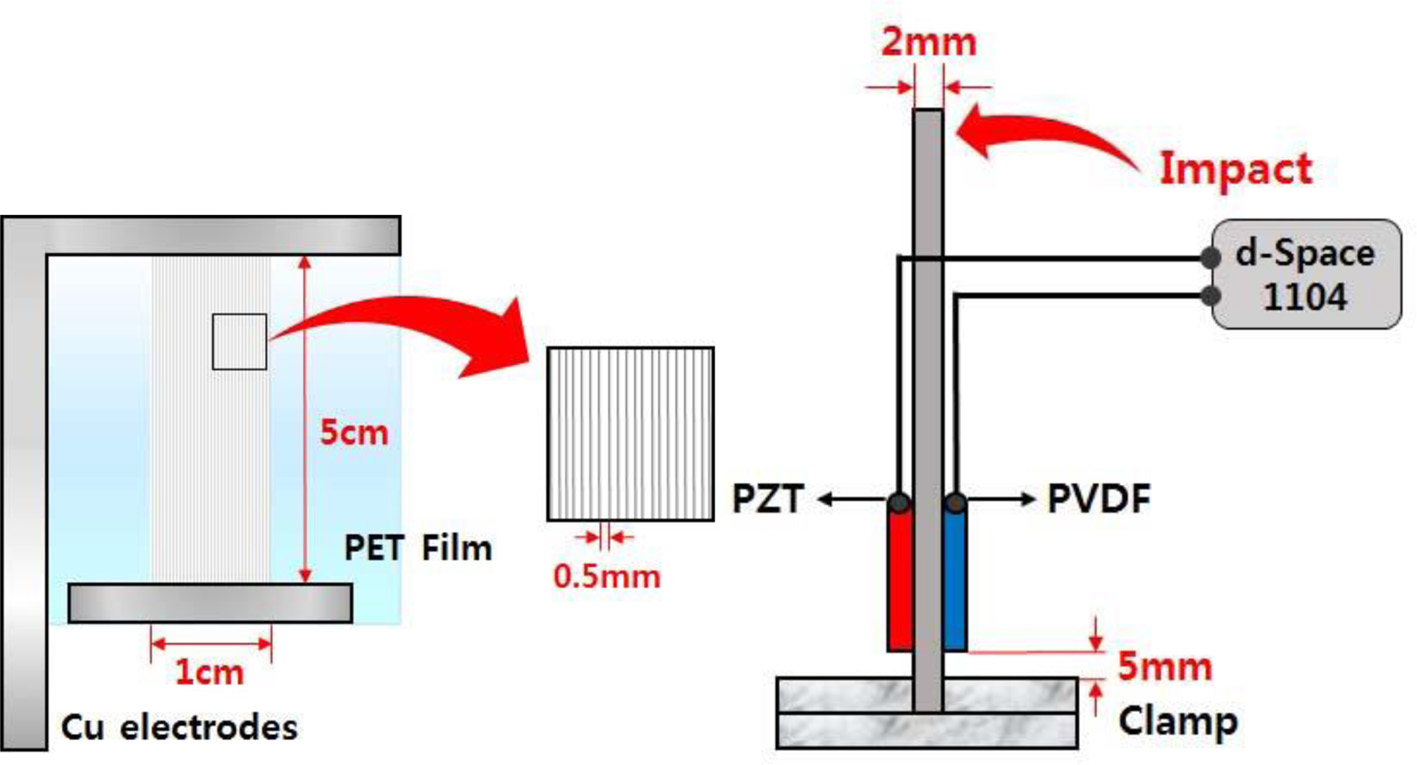
The 18 wt% poly(vinylidene fluoride) (PVDF) fibers were near-field electrospun at a flow rate of 139 nL/min, an electric field of 12 kV/cm, and a collector speed of 500 mm/s. A PVDF fiber array consisting of 20 fibers was adhered to the flexible PET film. A PVDF and a lead zirconate titanate (PZT) sensors were attached at a distance of 5 mm from the clamped end of the Al cantilever. The vibration sensing capabilities of sensors were examined by measuring the potential generated by the sensors during impact testing. Although the voltage of PVDF sensor was 200 times smaller than that of PZT sensor, the waveforms of the sensor output were similar. Both sensors were determined to be sensitive to variations in the level of dynamic strain due to the inherent piezoelectricity. The spectral results of both sensors exhibited the same signal generated by natural frequency of cantilever.
근거리 전기방사법을 이용하여 유량속도 139 nL/min, 전기장 세기 12 kV/cm, 콜렉터 속도 500 mm/s로 18 wt% 폴리(비닐리덴 플루오라이드)(PVDF) 섬유를 제조하였다. PVDF 섬유 모듈은 폴리(에틸렌 테레프탈레이트) 필름 위에 위치하였다. 모듈은 0.5 mm 간격으로 섬유 20줄로 구성되어 있다. 압전성과 진동 센서특성은 Al 외팔보를 이용한 충격실험을 통해 조사하였다. PVDF 센서와 lead zirconate titanate(PZT) 센서는 Al 외팔보 양면 끝에서 5 mm 위치에 에폭시를 이용 부착하였다. PVDF 센서의 전압특성은 PZT 센서보다 200배 정도 작았지만 출력 특성 파형은 유사하였다. 파장 측정 결과, 두 센서 모두 Al 외팔보의 고유 파장에서 관찰되었다.
A PVDF sensor consisting of 20 straight fiber lines was attached with epoxy at a distance of 5 mm from the clamped end of the Al cantilever. The cantilever size was 270×50×2 mm3 and the elastic modulus was 69 GPa. A commercial PZT sensor (27 mm piezo elements sounder sensor, P1113) was also attached to the opposite side of the same location. After attaching the sensors with the epoxy, the piezoelectric properties and vibration sensing capabilities of sensors were examined by measuring the electrical potential generated by the sensors during impact testing of the cantilever.
Keywords: poly(vinylidene fluoride), near-field electrospinning, lead zirconate titanate, sensor
Mechanical energy scavenging from human movements such as body movement and muscle stretching is an attractive renewable source of power for its applications ranging over nano-generator, tissue engineering, wearable sensors, nanodevices, and microelectromechanical systems.1-8 Zinc oxide(ZnO) nanowires were firstly used for nanogenerators due to the feasibility of utilizing inorganic nanomaterials with semiconducting and piezoelectric properties.5 Nowadays, the nanogenerators possessing nonsemiconducting and organic nanomaterials are emerging as the energy harvester due to direct integration with other structures and higher energy conversion efficiency.
Poly(vinylidene fluoride) (PVDF), a semi-crystalline polymer with -(CF2-CH2)- repeating units, has been considered as the material of choice for electrical devices and systems such as piezoelectric sensors, electro-mechanical actuators and energy harvesters because of controllable precision deposition of fibers, low cost, and easy operation.1-8 A flexible PVDF may
overcome the limitation of rigid ceramic sensors and can be used as a wearable sensor as well as a micro-energy harvester. PVDF exhibited high flexibility, minimizing resistance to external mechanical movements in low-frequency, largedeflection energy scavenging applications. In addition, PVDF has good piezoelectric, mechanical properties, and chemical stability. The use of PVDF active sensors is also suitable for the biomedical applications such as health monitoring methods due to their biocompatibilities and piezoelectric properties. The PVDF, a scaffold for bone tissue engineering, can generate electrical activity when mechanically deformed due to the presence of the piezoelectric β-phase.1
Among five different crystalline forms (α, β, ρ, δ, and ε) of PVDF polymer, the β crystal phase is the most important phase of PVDF. It is noted that the β crystal phase possessing the trans-type molecular chains provide the permanent dipoles arranged in the same direction, leading to large spontaneous polarization.1-8 The fabrication of the piezoelectric devices using electrospun PVDF fibers is a simple, direct and efficient way to obtain the β phase, mechanically stretched and electrically poled, from the α phase by means of near-field electrospinning (NFES).1-8 NFES can spin straight-line piezoelectric PVDF nanofibers on substrate with in situ mechanical stretching and electrical poling because of strong electric fields (≥ 107 V/m).2 In the present study, the parametric studies of PVDF nanofibers were investigated to optimize the NFES experimental conditions such as precursor solution concentration and collector moving speed (CMS). In addition, the piezoelectric properties and vibration sensing capabilities of PVDF sensors were examined by measuring the electrical potential generated by the sensors during impact testing of the Al cantilever.
Precursor Solution Preparation. PVDF (Mw=534000), dimethyl sulfoxide (DMSO), and capstone (FS-66) were purchased from Sigma-Aldrich. The precursor solution was prepared
from 2 g of PVDF in 6.329 mL of acetone (99.5%, Samchun, Korea) by stirring for 0.5 h. The capstone (0.5 g) dissolved in 4.546 mL of DMSO was then added to the above solution to obtain PVDF solution.6 The viscosity and the electrical conductivity of solutions were examined by using the viscometer (DV 1M, Brookfield, USA) with spindle NO. SC4- 31 at 6 rpm and the conductivity meter (Metrohm 912, Switzerland).7-9
Near-field Electrospinning (NFES). A 2-axis robot electrospinning apparatus (ESR200PR2, NanoNC, Korea) was used. A 30G B-D metal needle with an inner diameter of 0.14 mm and a 5 mL B-D luer-lok syringe were used to pump (KDS-200, Stoelting Co., USA) the precursor solution. The flow rate was 139 nL/min. A high voltage power supply (ES30P-5W, Gamma High Voltage Research Inc., USA) equipped with current and voltage digital meters was used, and
a voltage of 1.2 kV was applied across 1 mm of needle to ground collector distance. The desired patterning of the fibers was controlled by the coordinated movements of the x-y linear
stages (DTR2-44MS-SIG, Dasa Tech Co. Ltd, Korea) with a speed of 100~500 mm/s via Darwin pro (version 1.0.0) software. The PVDF fiber lines in the range of 1 to 100 were spun at intervals of 0.5 mm and the pattern was repeated up to 8 times. Crystal structure of PVDF fibers was then examined by Fourier transform infrared spectroscopy (FTIR, Spectrum Two, PerkinElmer, UK).
Characterization of Fiber. Piezoelectric transducers that are made of lead zirconate titanate (PZT) and PVDF have been extensively used in sensing, actuation, vibration control, and
harvesting energy from external movements.10 PVDF has also been characterized for use in pressure sensors, ultrasonic sensors, and for monitoring cardiorespiratory signal.10-12 A PVDF
sensor was attached with epoxy at a distance of 5 mm from the clamped end of the Al cantilever, as depicted in Figure 1. The cantilever size was 270×50×2 mm3 and the elastic modulus was 69 GPa.10 A commercial PZT sensor (27 mm piezo elements sounder sensor, P1113) was also attached to the opposite side of the same location. After attaching the sensors with the epoxy, the Al cantilever beam was kept for 24 h at room temperature. The piezoelectric properties and vibration sensing capabilities of sensors were examined by measuring the electrical potential generated by the sensors during impact testing of the cantilever. The Al cantilever beam was held firmly in a vertical direction on an anti-vibration table by using a clamp.
Time domain responses of the sensors located at a distance of 5 mm from the clamped end were measured when an impulse load was applied to the tip of the cantilever by using an impact hammer.10 The response of the Al cantilever beam was recorded during the impact test through the PZT sensor bonded at different location over the span of cantilever and the results
were compared to those obtained from PVDF sensors located at the opposite side of the same location. The output voltages of these sensors were collected using a 16-bit AD conversion
board (DS1104 R & D Controller Board, dSPACE GmbH) and were stored personal computer. The software for data acquisition was developed a real-time workshop of matlab & simulink
(Matlab, Matworks co.), throughout the experiment.10,13,14
|
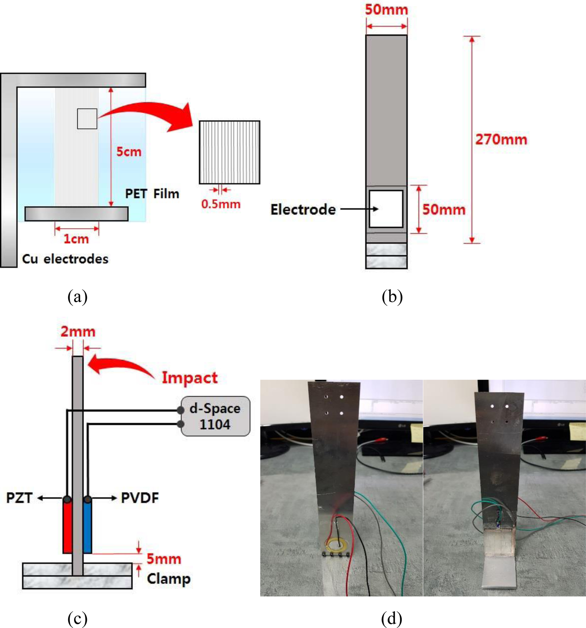
Figure 1 A schematic diagram of (a) PVDF fiber array; (b) front view; (c) side view of Al cantilever bonded with PZT and PVDF sensors. |
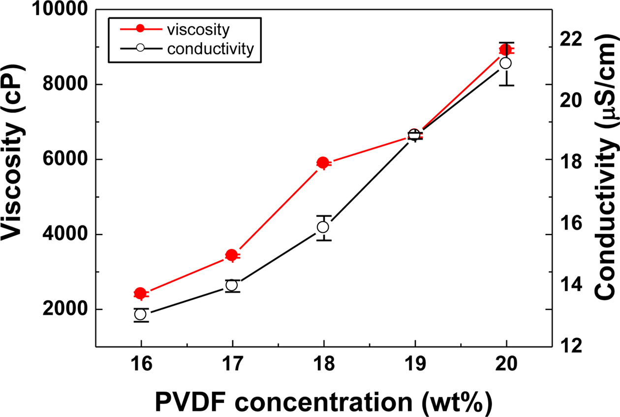
The viscosity and electrical conductivity of PVDF solution were shown in Figure 2. Viscosity provides the time to stay at the tip of nozzle during the electrospinning (ES). If the viscosity
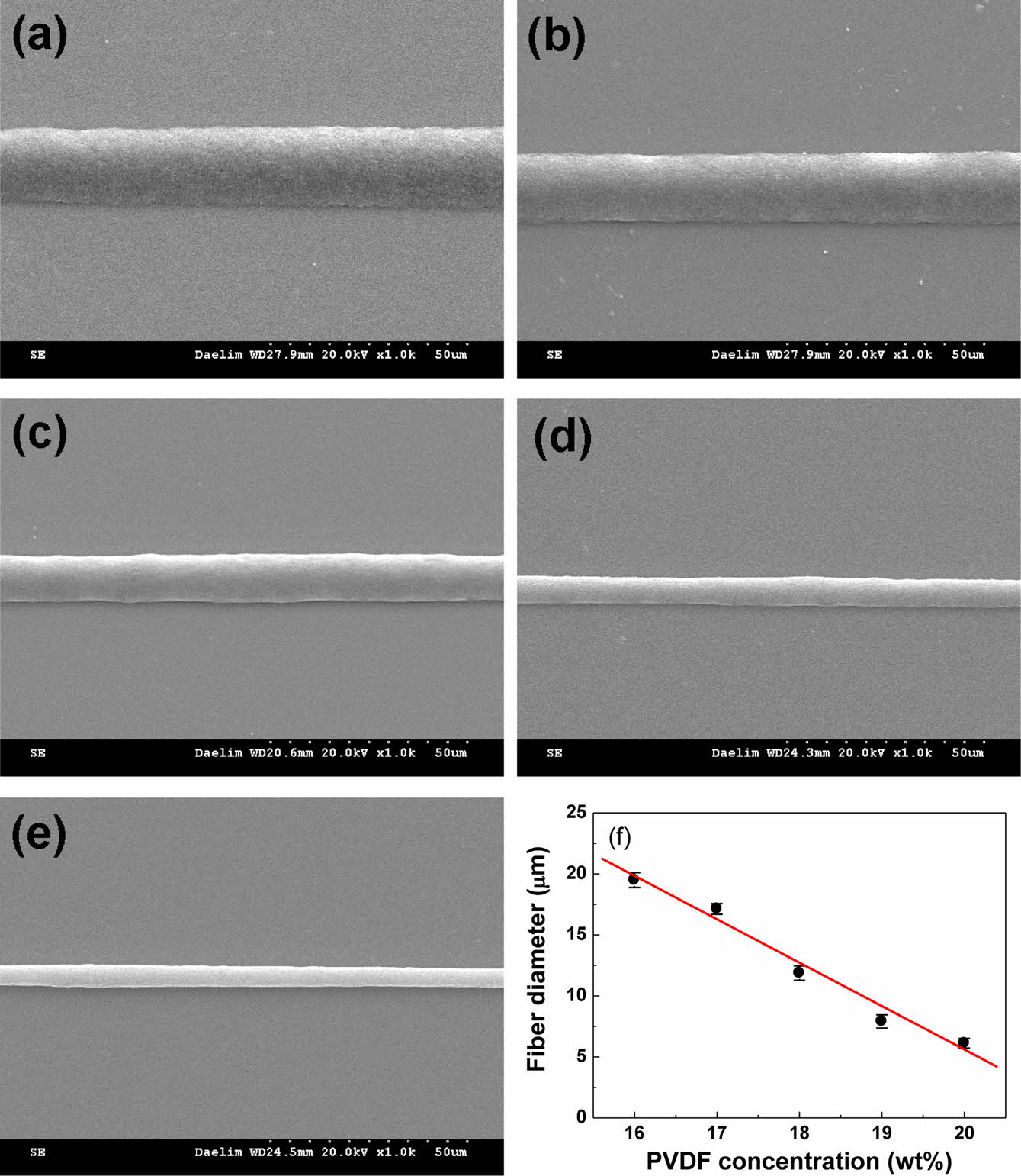
of the solution is too low or high, ES would not be possible. The viscosity and the conductivity of PVDF solution increased gradually from 2.4×10³ to 8.9×10³ cP, from 13 to 21 mS/cm with increasing the PVDF concentration from 16 to 20 wt%, respectively. The rheological results are in good agreement with the previous result.6 To determine the optimum combination of viscosity and conductivity, the morphology was examined. The morphology of PVDF fibers electrospun at a flow rate of 139 nL/min and an electric field of 12 kV/cm was studied as a function of polymer concentration, as depicted in Figure 3. It shows a typical SEM morphology of the straight-line-shape PVDF fibers. The fiber diameter decreased gradually from 19.4±0.6 to 6.1±0.3 μm with increasing the PVDF concentration from 16 to 20 wt%. It is reported that a high solution viscosity is preferred in order to limit the spreading of the as-deposited liquid fiber because most of the fiber solidification process takes place on the substrate.4 On the contrary, larger addition of PVDF may decrease the spinnability of the solution and make it unattainable to get continuous formation of fibers. Reasonably high PVDF concentration would be better to meet practical NFES needs through adjustment of viscosity and conductivity. This may give the reasoning behind 18 wt% of PVDF content chosen for the fabrication of the PVDF fibers.
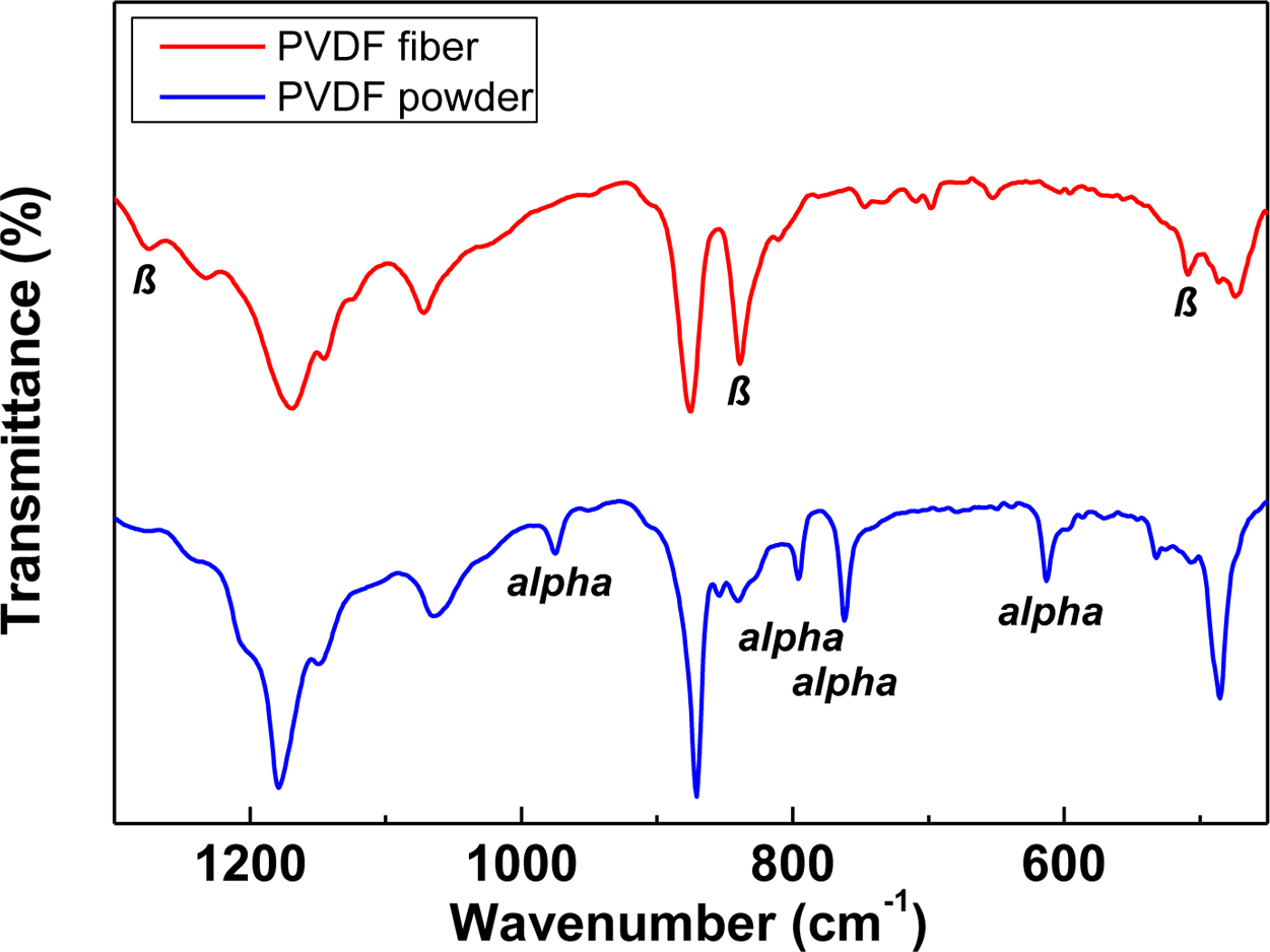
FTIR studies of PVDF powder and electrospun PVDF fibers are shown in Figure 4. Absorption of peaks of 510, 840 and 1278 cm-1 represent β crystal phase of PVDF. Absorption peaks of 614, 761, 796 and 976 cm−1 corresponded to α crystal phase, which is in good agreement with the results reported elsewhere.6 The PVDF powder exhibited a phase mixture of α and β, as displayed in Figure 4. However, absorption peaks of α phase were dramatically attenuated after NFES. On the other hand, the peaks corresponding to β phases became stronger after electrospinning compared to those of α phases, suggesting that NFES is an effective method for obtaining the β phase.
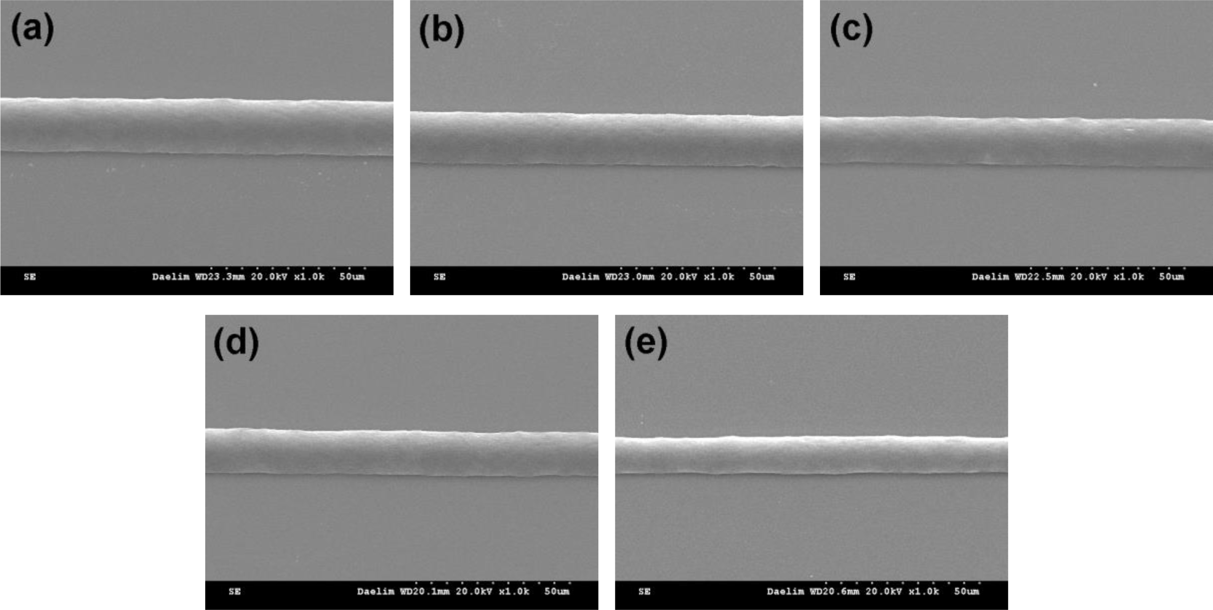
The straight-line-shape fibers were generated by controlling CMS in the range of 100 to 500 mm/s along a straight line. He et al. reported that the morphology of fibers was changed from
multicircle coil, single-circle coil, wavy line, to straight-line with increasing CMS from 50 to 400 mm/s.5 In the present study, the orderly direct-write fibers were obtained from a straight-line jet regardless of CMS (100~500 mm/s) probably due to the shorter spinning distance in NFES and the strong drag force from the moving collector. The diameter of the straight-line PVDF fibers decreased gradually from 17.5±0.3 to 11.8±0.4 μm with increasing CMS from 100 to 500 mm/s, as displayed in Figure 5. In the present study, a CMS of 500 mm/s was chosen for the fabrication of PVDF sensor via NFES.
The desired patterning of the PVDF fibers was controlled by the coordinated movements of the x-y linear stages with a speed of 500 mm/s via Darwin pro software. The PVDF fiber lines (1~100) were spun at intervals of 0.5 mm and the pattern was repeated up to 8 times. Stainless steel electrodes were used as the collector for NFES process. A PVDF fiber array was
spun on stainless steel substrate with a width of 80 mm and a length of 80 mm. The selected PVDF fiber array with a dimension of 10×50 mm was adhered to the flexible poly(ethylene
terephthalate) (PET) film to provide packaging protection. The array consists of 20 straight fibers 0.5 mm away from each other, as shown in Figure 1(a). Silver pastes were applied at the
two ends of PVDF fibers so that the bonding between fiber and copper electrodes are reinforced and the contact resistance is reduced.
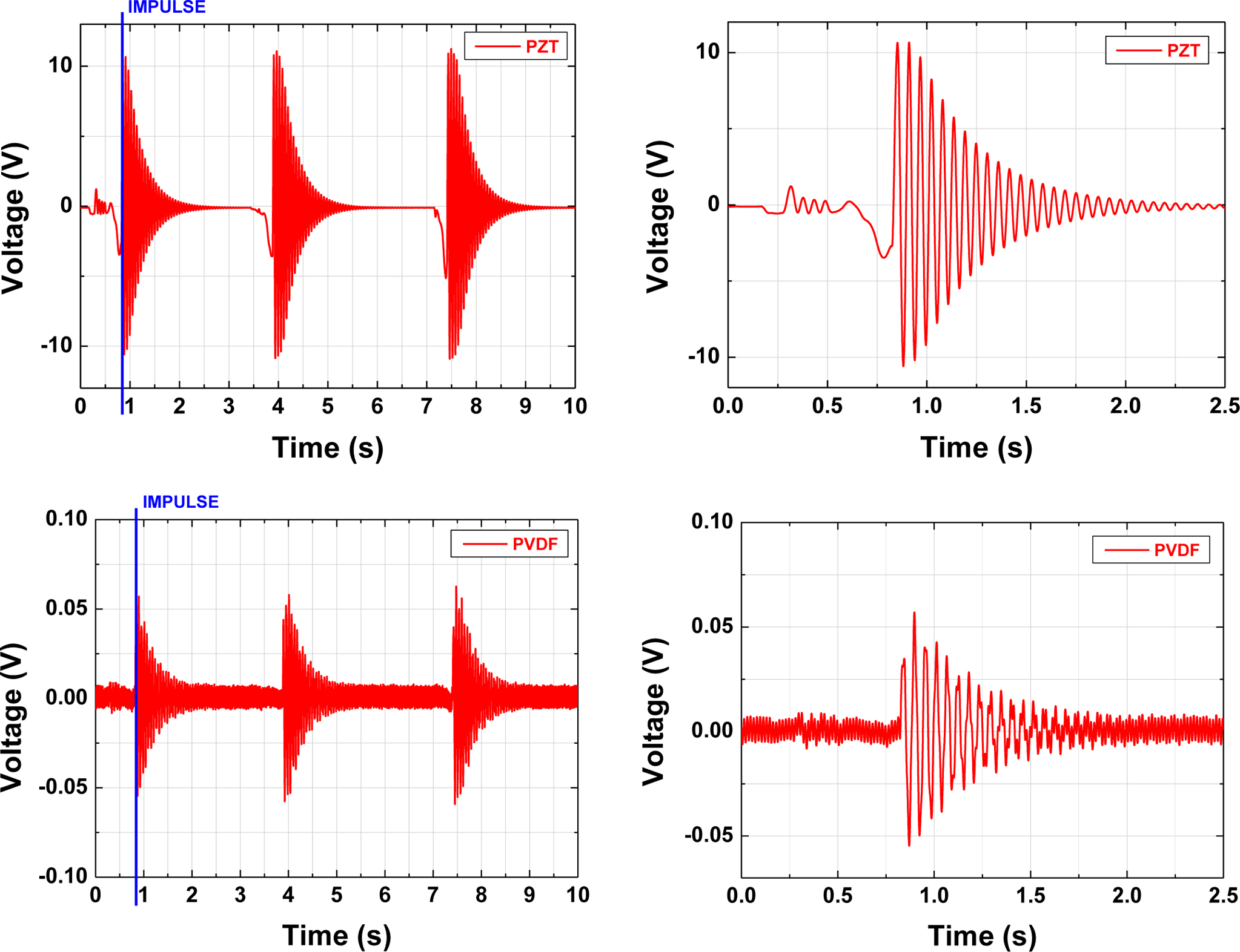
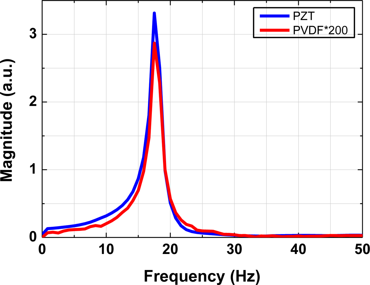
Time responses of the PVDF and PZT sensors when an impulse load was applied at the tip of the cantilever are shown in Figure 6. The sensing device can convert the mechanical energy of low-frequency ambient vibrations and impacts into electrical energy.8 The maximum output peak voltage of PVDF sensor (0.055 V) was about 200 times smaller than that (10.6 V) of PZT sensor. The waveform of the sensor output signal indicates that the vibration of the cantilever, mechanical energy, is well converted into an electric signal. In addition, the sensitivity of the PVDF sensor, represented by output voltage, can be improved by increasing the number of fibers on PVDF sensor. Both sensors were determined to be very sensitive to variations in the level of dynamic strain due to the inherent piezoelectricity.10 The spectral analysis was performed to evaluate the natural frequency of Al cantilever. Both PZT and PVDF sensor revealed that the signal was generated by the first-order natural frequency of the Al cantilever (~17 Hz), as displayed in Figure 7. The frequencies of two sensors coincided with each other, suggesting that the sensors can measure the resonant frequencies of vibrating structure accurately. Our previous studies revealed that PVDF was clinically safe and effective due to the absence of cytotoxicity and excellent cell proliferation.6
The increased demand for reliable and sensitive sensors that incorporate the well-known capabilities in such area as biomedical diagnosis has fueled the development and introduction
of flexible polymer-based sensors. The wearable biosensors have been actively researched by measuring the biological signals of human body. The human body contains three kinds of
muscle tissue (cardiac, smooth, and skeletal muscle) and each performs specific tasks to maintain homeostasis. Among muscles, the primary function of skeletal muscle is to convert
chemical energy to mechanical energy, resulting in muscle contraction.14-19 Contraction of skeletal muscle moves one part of the body with respect to another part, as in flexing the forearm. Contraction of several skeletal muscles in a coordinated manner moves the body in its environment, as in walking or swimming. A difficulty in preparing the textile-based sensor
using a conductive fabric in the form of fiber can be solved by transferring or attaching the flexible PVDF fiber sensor on textile.17 The battery of implantable devices such as artificial cardiac pacemaker and implantable cardioverter defibrillator have to be replaced every 10 years.18,19 The problem can be also worked out by using a flexible polymer-based generator.
Our previous studies revealed that the PVDF fibers exhibited cell viability of 98% compared to the negative control, as measured at a wavelength of 415 nm by using the microplate
absorbance spectrophotometer.6 The result of cell counting kit- 8 (CCK-8, Dojindo Molecular Technologies, Inc., Japan) cell proliferation test (measuring at a wavelength of 450 nm), L-
929 cells adhered well to PVDF and proliferated continuously (0→20%→28%→58%→98%) with increasing time (0→6 h→12 h→24 h→48 h), indicating that the PVDF fibers are likely to be highly applicable to the biomedical applications due to their piezoelectric, mechanical properties, and biocompatibilities.
|
Figure 2 The variation of electrical conductivity and solution viscosity |
|
Figure 3 SEM images of PVDF fibers with different PVDF concentrations: |
|
Figure 4 FTIR spectra of PVDF powder and PVDF fiber. |
|
Figure 5 SEM images of the electrospun PVDF fibers at various collector moving speed; (a) 100 mm/s; (b) 200 mm/s; (c) 300 mm/s; (d) |
|
Figure 6 Time responses of PZT and PVDF sensors bonded on Al cantilever beam by applying an impulse load at the tip of the cantilever. |
|
Figure 7 Power spectral density curves of PZT and PVDF sensors. |
PVDF fibers in the composition range of 16 to 20 wt% were electrospun by means of NFES at a flow rate of 139 nL/min and an electric field of 12 kV/cm. The PVDF fibers were optimized
by examining the parameters of PVDF composition and CMS. The 18 wt% PVDF fibers were finally spun at a flow rate of 139 nL/min, an electric field of 12 kV/cm, and CMS of 500 mm/s to fabricate the electrical transducer. A selected PVDF fiber array with a dimension of 10×50 mm was prepared and the array consisted of 20 straight fibers 0.5 mm away from each other. The piezoelectric properties and vibration sensing capabilities of sensors were measured by detecting the electrical potential generated by the sensors during impact testing of the Al cantilever. Although the voltage of PVDF sensor was about 200 times smaller than that of PZT sensor, the waveform of the sensor signal was similar. Both sensors were determined to be very sensitive to variations in the level of dynamic strain due to the inherent piezoelectricity. The spectral analysis results of both sensors, exhibiting the same signal generated by natural frequency of Al cantilever, suggested that PVDF fibers can be highly applicable to biomedical applications such as health monitoring sensors (monitoring cardiorespiratory signal) and generators.
- 1. P. Calvert, Nature, 256, 694 (1975).
-

- 2. C. Chang, V. H. Tran, J. Wang, Y. Fuh, and L. Lin, Nano Lett., 10, 726 (2010).
-
- 3. D. Sun, C. Chang, S. Li, and L. Lin, Nano Lett., 6, 839 (2006).
-
- 4. Y. Y. S. Huang, E. M. Terentjev, T. Oppenheim, S. P. Lacour, and M. E. Welland, Nanotechnology, 23, 105305 (2012).
-
- 5. X. He, J. Zheng, G. Yu, M. You, M. Yu, X. Ning, and Y. Long, J. Phys. Chem., 121, 8663 (2017).
-
- 6. J. Rho, D. Y. Lee, M. Lee, B. Kim, and H. Jeong, J. Korean Cryst. Growth Cryst. Technol., 28, 1 (2018).
- 7. Y. Wang, K. Ren, and Q. M. Zhang, Appl. Phys. Lett., 91, 222905 (2007).
-
- 8. Z. H. Liu, C. T. Pan, C. Y. Su, L. W. Lin, Y. J. Chen, and J. S. Tsai, Sens. Actuator A, 211, 78 (2014).
-
- 9. G. Oh, J. Rho, D. Y. Lee, M. Lee, and Y. Kim, Macromol. Res., 26, 48 (2018).
-
- 10. Z. Abas, D. H. Yang, H. S. Kim, M. K. Kwak, and J. Kim, Intl. J. Appl. Mech., 7, 1550065 (2015).
-
- 11. A. V. Shirinov and W. K. Schomburg, Sens. Actuators A, 142, 48 (2008).
-
- 12. V. T. Rathod, D. R. Mahapatra, A. Jain, and A. Gayathri, Sens. Actuators A, 163, 164 (2010).
-
- 13. D. Y. Lee, M. Lee, K. J. Kim, S. Heo, B. Kim, and S. Lee, Surf. Coat. Technol., 200, 1920 (2005).
-
- 14. D. Y. Lee, I. Park, M. Lee, K. J. Kim, and S. Heo, Sens. Actuators A, 133, 117 (2007).
-
- 15. D. Y. Lee, J. Cho, Y. Kim, and Y. Oh, J. Korean Sens. Soc., 17, 281 (2008).
-
- 16. D. Y. Lee, Y. Kim, S. Lee, M. Lee, J. Lee, B. Kim, and N. Cho, Mater. Sci. Eng. C, 28, 294 (2008).
-
- 17. M. Stoppa and A. Chiolerio, Sens., 14, 11957 (2014).
-
- 18. G. Hwang, H. Park, J. Lee, S. Oh, K. Park, M. Byun, H. Park, G. Ahn, C. K. Jeong, K. No, H. Kwon, S. Lee, B. Joung, and K. J. Lee, Adv. Mater., 26, 4880 (2014).
-
- 19. Y. Qi and M. C. McAlphine, Energy Environ. Sci., 3, 1275 (2010).
-

- Polymer(Korea) 폴리머
- Frequency : Bimonthly(odd)
ISSN 0379-153X(Print)
ISSN 2234-8077(Online)
Abbr. Polym. Korea - 2023 Impact Factor : 0.4
- Indexed in SCIE
 This Article
-
2019; 43(5): 764-770
Published online Sep 25, 2019
- 10.7317/pk.2019.43.5.764
- Received on May 17, 2019
- Revised on Jun 18, 2019
- Accepted on Jun 27, 2019
Services
Shared
Correspondence to
- Deuk Yong Lee
-
Department of Biomedical Engineering, Daelim University, Anyang 13916, Korea
- E-mail: dylee@daelim.ac.kr
- ORCID:
0000-0003-1674-412X
Hyecheon Building(Room 601), #354, Gangnam-Daero, Gangnam-Gu, Seoul 06242, Korea
TEL : 82-2-568-3860, 561-5203, 569-3860 FAX : 82-2553-6938 E-mail: polymer@polymer.or.kr